How to Improve Results with HCP Antibody Coverage Techniques
In this post, I want to share what I’ve learned through firsthand experience—actionable steps that can help you optimize your HCP antibody coverage techniques for better, cleaner, and more compliant results.
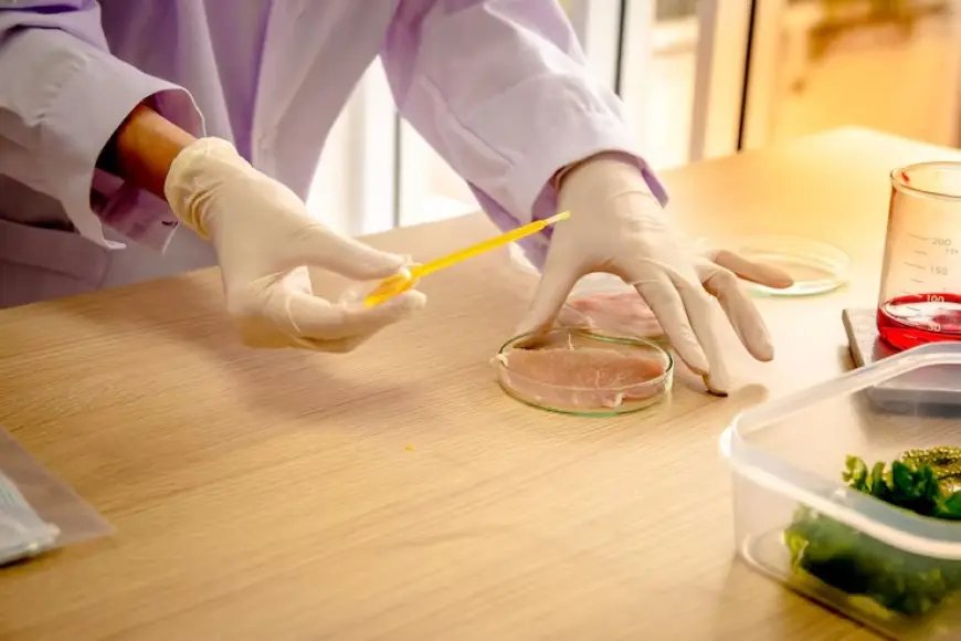
When I first began working with host cell protein (HCP) detection in biologics, I quickly realized that antibody coverage is one of the most critical yet challenging aspects to get right. As a scientist striving for precise and reliable biologics development, understanding how to improve HCP antibody coverage has not only improved my assay sensitivity but also enhanced my compliance with regulatory expectations.
In this post, I want to share what I’ve learned through firsthand experience—actionable steps that can help you optimize your HCP antibody coverage techniques for better, cleaner, and more compliant results.
Understanding the Core of HCP Antibody Coverage
Host cell proteins are byproducts from expression systems like E. coli, CHO cells, or yeast that can potentially contaminate your biologic product. To detect and quantify these proteins, we rely heavily on polyclonal antibodies that recognize a wide range of HCPs.
But here’s the catch—not all antibodies offer the same coverage. Inadequate antibody coverage can lead to misleading data and regulatory setbacks. If you're wondering why your ELISA isn't giving expected results, poor antibody coverage may be the culprit.
Improving antibody coverage is not about using more antibodies—it’s about using the right combination, developed and validated through effective techniques.
Step 1: Selecting the Right Host Cell System
It all starts with choosing the correct host cell system. Each system produces a different array of HCPs, which means the antibodies developed for one system won’t always work well with another.
In my experience, working with CHO cell lines required a completely different antibody pool than what was needed for E. coli-based expression. Don’t assume cross-compatibility. Perform a detailed proteomic profile of your cell line first to determine the types of HCPs present.
Step 2: Using 2D Western Blot for Antibody Evaluation
The turning point in my antibody coverage journey came when I began using 2D Western Blot analysis.
Unlike traditional 1D methods, the 2D approach allowed me to visualize how many HCPs were being detected by my polyclonal antibodies. It helped me assess coverage comprehensively. I could directly overlay the blot image with silver-stained 2D gels and determine which proteins were missed.
When done consistently, this method offers both qualitative and quantitative insights into how effective your antibodies are.
Step 3: Leveraging Mass Spectrometry
Mass spectrometry has become an indispensable tool in my lab. Once we identify which proteins are not being detected by ELISA, we use MS to analyze those gaps.
This step was critical during my work with biosimilar development. Regulatory authorities expected not just detection, but explanation. MS gave us the power to map out the full protein profile, even those undetected by antibodies, helping us refine our strategy and develop targeted antibodies for critical HCPs.
This deep dive into proteomic characterization helped us meet stringent expectations, and I highly recommend integrating MS into your workflow if you haven’t already.
Step 4: Pooling and Affinity Purification
Another effective method I’ve employed is pooling antibodies raised against various antigen preparations. This includes both whole cell lysates and purified protein mixtures.
We didn’t stop there. We enhanced the quality by using affinity purification to remove non-specific antibodies, increasing the specificity of our HCP ELISA. By combining pools, we increased the breadth of HCP detection, which significantly improved our assay’s performance.
One tip—ensure each antibody batch is well-characterized. Pooling should not be a shortcut for quality, but rather a refinement process to enhance coverage.
Step 5: Testing for Cross-Reactivity and Redundancy
Not all antibody improvements are about expansion—sometimes it's about refinement. I’ve made the mistake of using a pool that resulted in high background noise due to cross-reactivity. It was a painful lesson in how too much redundancy can mask important signals.
To resolve this, I started validating each antibody pool not just for coverage but also for specificity and reproducibility. Running parallel ELISAs and testing various batches under different stress conditions allowed me to understand antibody behavior across scenarios.
Reducing cross-reactivity while maintaining coverage is a balancing act—but one that’s essential for long-term assay reliability.
Step 6: Regulatory Expectation Alignment
The FDA and EMA both emphasize robust characterization of HCP antibodies. Your coverage data must stand up to regulatory scrutiny.
I once worked on a project where our 2D Western Blot showed 65% antibody coverage—acceptable in many settings—but regulators flagged the unknowns in the remaining 35%. We went back to the lab, used MS to identify them, and found that some were immunogenic.
That experience taught me to proactively map and disclose all detectable HCPs, with justification for those that remain undetected. Regulators value transparency and scientific rationale more than perfect numbers.
Step 7: Continuous Improvement Through Risk Assessment
Even after assay validation, I conduct regular risk assessments as part of process development. Over time, expression systems evolve, and so do HCP profiles.
Monitoring batch-to-batch variation has helped me spot antibody performance drift early on. It’s not enough to verify coverage once—it's an ongoing responsibility, especially when scaling up.
I strongly recommend setting a schedule for reassessing antibody coverage at key milestones—like cell line changes, purification process updates, or scale-up stages.
A Reliable Partner in the Process
I’ve had the privilege of working with service providers who offer detailed antibody coverage assessments. These collaborations saved us months of in-house development time. Whether you outsource or do it internally, the key is maintaining rigorous documentation, transparency, and scientific integrity throughout.
For those still figuring out the best route, I suggest Going Here to learn more about providers and platforms that offer robust antibody coverage solutions tailored to your cell line and regulatory needs.
Final Thoughts: Precision, Patience, and Process
Improving HCP antibody coverage is not a one-off task—it’s a strategic and ongoing investment. In my experience, focusing on data integrity, antibody specificity, and a strong understanding of your HCP landscape can transform your biologic development process.
Whether you're just getting started or fine-tuning your ELISA, I hope these insights will guide you to more reliable and reproducible results. Keep pushing forward. Each improvement in antibody coverage brings you closer to safer, more effective therapeutics—and that’s the ultimate goal we all share in this industry.
For more insights and services in optimizing HCP antibody strategies, Click This Link to explore current tools and technologies shaping the future of biologics.
Need expert support to enhance your HCP strategy? Contact us today to get personalized guidance and proven solutions.
What's Your Reaction?
 Like
0
 Dislike
0
 Love
0
 Funny
0
 Angry
0
 Sad
0
Sad
0
 Wow
0
Wow
0


