Nuclear Cardiology: How Advanced Imaging is Revolutionizing Heart Health
 izan
izan

In the evolving field of cardiology, Nuclear Cardiology stands out as a cutting-edge diagnostic tool that enhances the detection and treatment of heart diseases. By using specialized imaging techniques and radioactive tracers, this branch of cardiology provides crucial insights into heart function, blood flow, and potential abnormalities. As cardiovascular diseases continue to be a leading cause of mortality worldwide, the advancements in Nuclear Cardiology are proving to be life-saving.
What is Nuclear Cardiology?
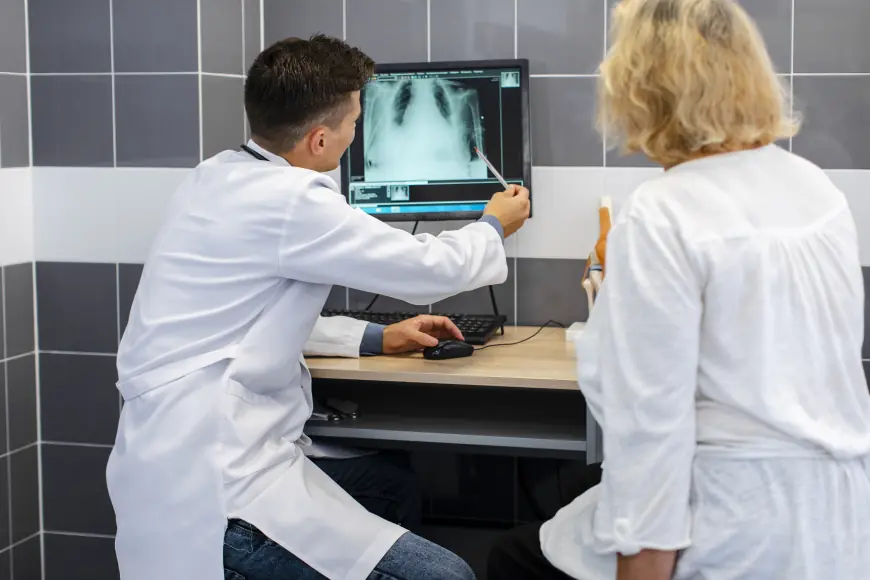
Nuclear Cardiology is a subspecialty of cardiology that utilizes non-invasive imaging techniques to assess the health and functionality of the heart. By injecting small amounts of radioactive tracers into the bloodstream, nuclear imaging allows doctors to evaluate blood flow, detect blockages, and analyze heart muscle performance with high precision.
Unlike conventional imaging methods, such as echocardiography or CT scans, nuclear imaging provides functional rather than just structural information. This makes Nuclear Cardiology particularly valuable in diagnosing coronary artery disease (CAD), assessing heart attack damage, and planning treatment strategies for various cardiac conditions.
How Nuclear Cardiology Works
The process of Nuclear Cardiology involves several steps, each contributing to a comprehensive heart health assessment:
-
Administration of Radioactive Tracers: A small amount of a radioactive substance, such as Technetium-99m or Thallium-201, is injected into the patient’s bloodstream. These tracers travel to the heart and emit gamma rays, which can be captured by specialized imaging cameras.
-
Imaging Techniques: Two primary imaging techniques are used in Nuclear Cardiology:
-
Single Photon Emission Computed Tomography (SPECT): Captures 3D images of the heart to analyze blood flow and detect areas with reduced perfusion.
-
Positron Emission Tomography (PET): Provides high-resolution images with superior accuracy, often used for evaluating heart tissue viability and detecting ischemic areas.
-
-
Stress and Rest Phases: The imaging process is usually conducted in two phases:
-
Stress Imaging: The patient undergoes physical exercise or is given a pharmacological agent (such as adenosine or dobutamine) to simulate exertion, helping identify areas of inadequate blood supply.
-
Rest Imaging: The heart is imaged in a relaxed state to compare results and assess overall function.
-
-
Image Analysis: Cardiologists interpret the nuclear images to detect any irregularities in blood flow, heart muscle viability, or previous cardiac damage. This data helps in diagnosing conditions and guiding treatment plans.
Applications of Nuclear Cardiology
1. Diagnosing Coronary Artery Disease (CAD)
One of the most critical uses of Nuclear Cardiology is in detecting CAD, which occurs when arteries supplying blood to the heart become narrowed or blocked. A nuclear stress test can identify regions of the heart that are not receiving sufficient blood, helping doctors determine the severity of the disease and plan interventions like angioplasty or bypass surgery.
2. Assessing Myocardial Viability
After a heart attack, it is crucial to determine whether parts of the heart muscle have survived or suffered permanent damage. Nuclear Cardiology helps differentiate between viable and non-viable tissue, allowing doctors to recommend the best course of treatment, including revascularization procedures or lifestyle modifications.
3. Evaluating Heart Function (Ejection Fraction Analysis)
The ejection fraction (EF) measures the percentage of blood pumped out of the heart with each beat. A nuclear scan provides precise EF values, which are essential for diagnosing and monitoring conditions like heart failure and cardiomyopathy.
4. Monitoring Post-Treatment Progress
For patients who have undergone cardiac procedures such as stenting or bypass surgery, Nuclear Cardiology plays a crucial role in monitoring the effectiveness of treatment and ensuring optimal heart function over time.
Advantages of Nuclear Cardiology
High Sensitivity and Accuracy
Compared to other imaging modalities, Nuclear Cardiology provides highly accurate functional data, enabling early detection of heart disease before symptoms appear.
Non-Invasive and Safe
While radiation exposure is involved, the amounts used are minimal and generally safe for patients. The non-invasive nature of the tests makes them suitable for routine screening and follow-up assessments.
Customized Treatment Plans
By precisely identifying the severity and location of heart disease, nuclear imaging helps cardiologists tailor treatments to individual patients, ensuring better outcomes.
Limitations and Considerations
Despite its benefits, Nuclear Cardiology has certain limitations:
-
Radiation Exposure: Although minimal, it may not be suitable for pregnant women or individuals with contraindications to radiation.
-
Time-Consuming: The process, particularly stress testing, can take several hours to complete.
-
Cost: Compared to other imaging techniques, nuclear scans can be expensive, potentially limiting accessibility for some patients.
The Future of Nuclear Cardiology
As technology continues to evolve, Nuclear Cardiology is expected to see significant advancements, such as:
-
Artificial Intelligence (AI): AI-driven image analysis is improving diagnostic accuracy and reducing interpretation times.
-
New Radiotracers: Ongoing research is focused on developing safer and more effective radiotracers with reduced radiation exposure.
-
Hybrid Imaging: Combining nuclear imaging with CT or MRI is enhancing diagnostic capabilities by providing both anatomical and functional insights.
Conclusion
Nuclear Cardiology is transforming the way heart diseases are diagnosed and managed. By offering unparalleled insights into blood flow, heart function, and tissue viability, it plays a crucial role in early detection, treatment planning, and long-term cardiac care. While there are some limitations, the benefits of this advanced imaging technique far outweigh the drawbacks, making it an indispensable tool in modern cardiology.
For those at risk of heart disease, undergoing a Nuclear Cardiology evaluation can be a life-saving decision. If you or a loved one is experiencing symptoms such as chest pain, shortness of breath, or fatigue, consult a cardiologist to determine if a nuclear imaging test is right for you.
With ongoing advancements in imaging technology, the future of Nuclear Cardiology looks promising, paving the way for more precise, efficient, and personalized heart care solutions.